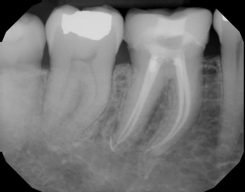
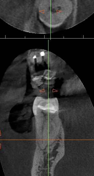
Honestly, this 2D PA didn’t look that suspicious to me.
🙈
Truly!
🤷♀️
I honestly wouldn’t have suspected a radix behind here!
🤔
Why does this matter??
In the literature the frequency of Radix Entomolaris can be as high as 30%!!!!
Usually on the DLs of mandibular first molars
😬
2nd screenshot abstract article by Calberson, et al. JOE 2007 shares that we should remember and appreciate how tricky these cases are.
On my very 1st radix ever I separated a file.
💔😔
It happens to almost every endodontist I’ve spoken with, especially when you don’t realize (without 3D imaging) that you have a radix (can sometimes appreciate it on the post op Xray).
To say that I fear these is an understatement.
Pearl: Use CBCT for endo!
Even 3 years ago, I wouldn’t have taken a #cbct for a lower molar for primary (non-retx) endo… I’ve done plenty of lower molar endos without cbct…. But, now, I am SO glad I have this technology and I take FULL advantage of it. I never regret taking a scan.
Without my CBCT imaging, I would have gone into this tooth without as much caution, and might have separated an instrument (like I did on my first radix). I am thankful the CBCT scan provided me the info I needed to avoid any mishaps.
I am extremely cautious and practically hold my breath during the entire procedure when it comes to honoring these severe radix curves.
And, my final PA also looks like this tooth is more benign than it was – radix, calcified, through a crown, and it was a HOT tooth tooooooo!
Trifecta +++++


